
Confession time here: microscopes are one of my favourite things, ever! I love investigating things with a microscope and could happily spend hours messing about with one. The strangest experiment I ever did was to get a friend’s cat to lick a microscope slide so we could see what was in her mouth. In this post, I will talk about how scientists use the latest in microscope technology to study viruses.
The use of lenses to make objects look bigger dates back to the 13th century. It’s not certain who actually invented the microscope; but they started to appear in the mid 17th century. Italian scientist Galileo Galilei played an important part, using his knowledge of telescopes to improve on earlier inventions.
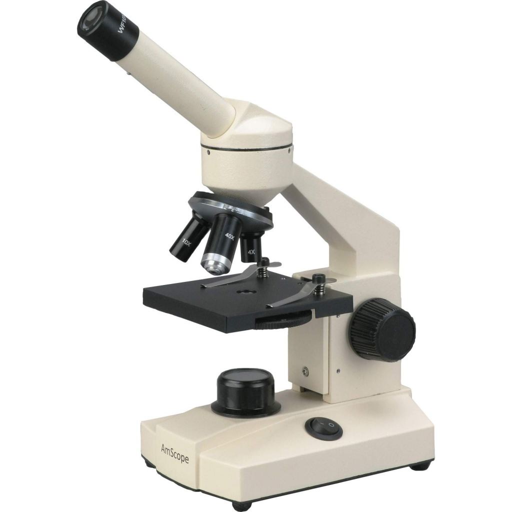
If you’ve used a microscope at school, it will have been an optical microscope, also sometimes called a light microscope. If you haven’t used one yet, you will do this in Year 7 and trust me, it’s amazing! Optical microscopes work by shining a light through a sample, and using a combination of lenses to magnify the image. Often, a stain is used – for example, methylene blue to stain animal/ human cells; or iodine for plant cells. Sometimes a combination is used – for example, Wright’s stain, used to examine blood, is a mixture of methylene blue which stains white blood cells, and a different stain for red blood cells.

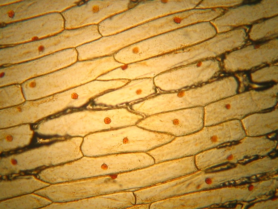
Optical microscopes have some limitations. The first is magnification. Mitochondria (the organelles of a cell that release energy) are the smallest things that can be seen with a standard optical microscope. Advanced computer processing does allow smaller objects to be observed; but this is very specialised, and still doesn’t produce the magnification needed to see viruses.
The second limitation of an optical microscope is that the images are two-dimensional – in other words, everything appears to be flat. Cells, like the ones seen above, are not flat but are three-dimensional. Being able to study cells, viruses and bacteria in 3-D is very important if we want to properly understand their structure. So, instead of optical microscopes, scientists studying COVID-19 and other viruses will be using electron microscopes.

Optical microscopes work by shining light through a sample. Electron microscopes use a beam of electrons instead of light. This has two advantages. First, you can achieve much greater magnification and resolution (resolution is how detailed and clear an image is); second, an electron microscope can give 3-D images. The disadvantages are that they are expensive; they are not portable; and some samples require very specialised preparation.

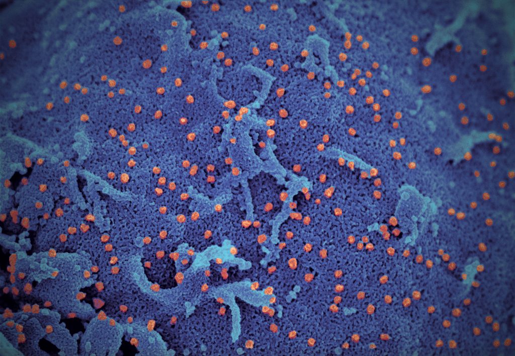
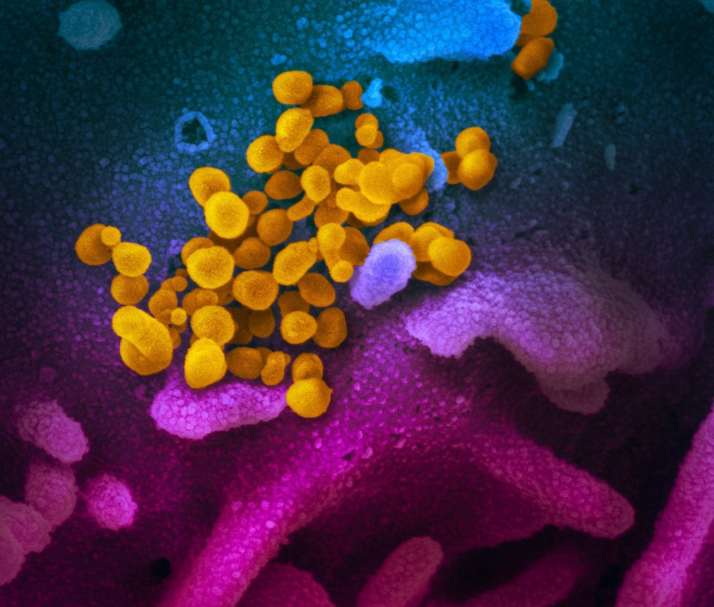
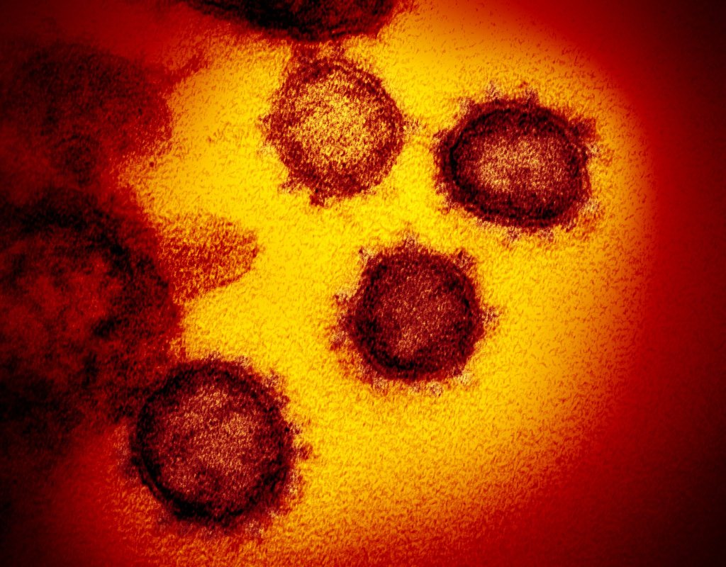
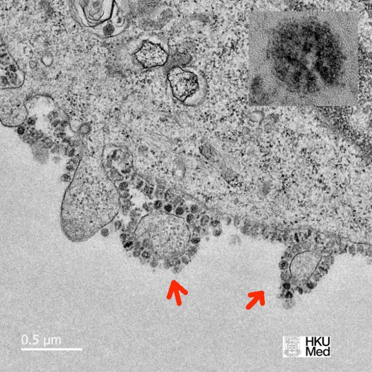
So, how will scientists be using microscopes to study the virus? There are two types of electron microscopy: scanning electron microscopy (SEM) and transmission electron microscopy (TEM). They can both do different things but broadly speaking, SEM is used to study objects in 3-D and TEM in 2-D.
SEM is particularly useful for scientists studying the structure of the virus, and how the virus attaches to the surfaces of human cells. TEM is very useful for studying how the virus behaves inside infected cells. Some pictures are shown below.

I hope you have found this interesting! In my next post, I will explain the technique scientists use to study proteins and other molecules on the surface of the virus: crystallography.
